
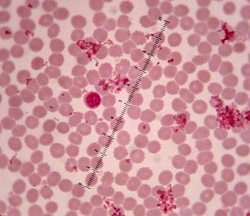
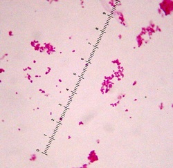
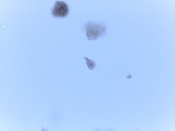
Slide 3 Bacteria and blood. The large cell near the middle is a lymphocyte; clusters of cocci are scattered among the red blood cells. Why are the bacteria so much smaller than the blood cells?
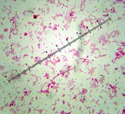
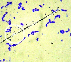
Slide 4 Pseudomonas aeruginosa (gram stain) These are gram negative rods
Slide 5 Bacillus subtilis with endospores. If you look carefully, you can see the clear areas inside some cells where the endospores are. Endospores do not take up a stain well.
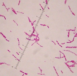
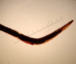
Slide 6 Vibrio fischeri (simple stain)- Note the comma-shaped cells in the center of the picture.
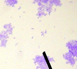
Slide 7 Neisseria gonorhoeae (gram stain) These are gram negative
diplococcic.
diplococcic.
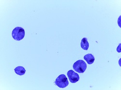
Slide 8 Streptococcus pneumoniae. These cells typically form pairs of cocci (diplococci); note the 3 pairs in the white circle.
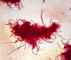
Slide 11 Borrelia burgdorferi - spirochetes
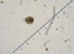
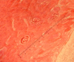
Slide 12 Entamoeba histolytica trophozoites (1000X) Note the prominent nucleus with chromatin thickening on the periphery
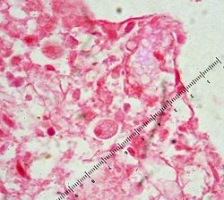
Slide 12 Entamoeba histolytica, (in situ - 1000X) This is a
cross-section of small intestine. Note the parasite just above the
ruler. Can you find the other one?
cross-section of small intestine. Note the parasite just above the
ruler. Can you find the other one?
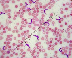
Slide 13 Trypanosoma gambiense, (blood smear-1000X)
Note the distinctive undulating membrane.
Note the distinctive undulating membrane.
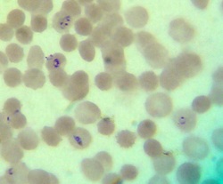
Slide 14 Plasmodium sp. (blood smear, 1000X) Note the typical signet ring-shaped trophozoites in the red blood cell on the far right. Some of the red blood cells are multiply-infected.
Slide 15 Taenia pisiformis proglottid. The pointer is on the ovary; testes are present but not visible (tapeworms are hermaphroditic).
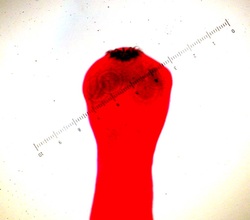
Slide 15 Taenia pisiformis, (40X) scolex with attachment hooks and
suckers.
suckers.
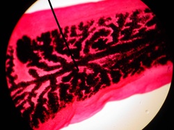
Slide 15 Taenia pisiformis, gravid proglottid. This segment shows the ovary branches filled with eggs.
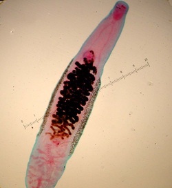
Slide 16 Clinorchis sinensis This is a fluke. Note the typical
leaf-shapted body form. There is an attachment sucker at the anterior end and another one just anterior to the dark, egg-filled uterus. The testes are the pink structures at the posterior end.
leaf-shapted body form. There is an attachment sucker at the anterior end and another one just anterior to the dark, egg-filled uterus. The testes are the pink structures at the posterior end.
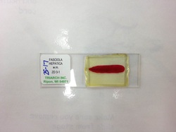
Slide 17 Fasciola hepatica
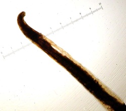
Slide 18 Necator americanus (hookworm) Note the hook shape at the anterior end.
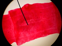
Slide 19 Trichinella spiralis (in situ) Note the encysted larvae (above the ruler) in these muscle fibers.
Slide 20 Enterobius vermicularis (pinworm).
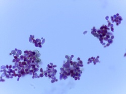
Slide 21 Saccharomyces sp., budding.
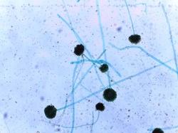
Slide 22 Aspergillus sp., conidia
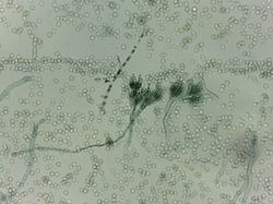
Slide 23 Penicillium sp., conidia
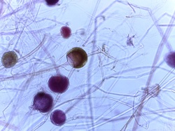
Slide 24 Rhizopus stolonifera. Sporangia.
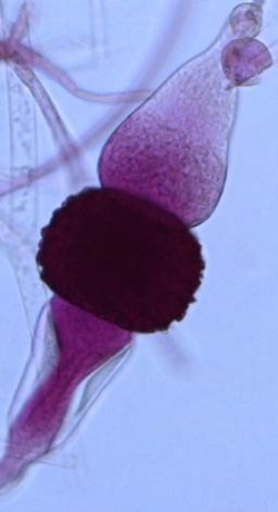
Slide 24 Rhizopus stolonifera. Zygospores.
Slide 28 Giardia lamblia, trophozoid stage
Slide 29 Trichomonas vaginalis
